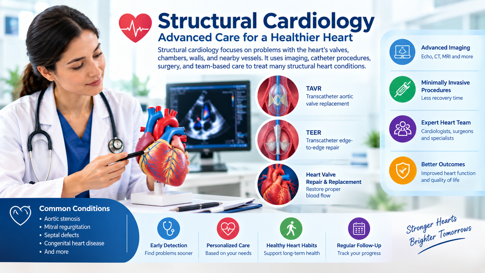

Structural cardiology is a heart field focused on problems in the heart’s valves, chambers, walls, and nearby vessels. It uses imaging, catheter procedures, surgery, and team-based care to treat many structural heart problems.
Have you ever wondered why a doctor checks your heart valves so closely?
Your heart has four chambers and four valves.
These parts work together to move blood through your body.
A problem with one part can change how your heart works.
Structural cardiology focuses on these physical heart problems.
It covers many conditions that affect heart shape or blood flow.
Some problems start before birth.
Others develop with age or other heart conditions.
Modern care can use medicines, surgery, or catheter procedures.
A catheter is a thin tube used inside blood vessels.
Doctors may guide it toward the heart during treatment.
Patients may also need several types of heart imaging.
These tests help doctors see the heart and its valves.
The care team then studies the results together.
This guide explains structural heart disease in simple terms.
You will learn common types, symptoms, tests, and treatments.
We also cover careers, fellowship training, and salary information.
What Is Structural Cardiology?
Structural cardiology deals with physical problems inside or around the heart.
These problems can involve valves, chambers, walls, or major blood vessels.
The heart has four chambers.
The upper chambers are called atria.
The lower chambers are called ventricles.
Four valves control blood flow through these chambers.
They open and close with each heartbeat.
A healthy valve helps blood move in one direction.
Structural heart doctors treat problems affecting these parts.
They may also treat some birth defects.
These defects can change the heart’s shape or blood flow.
Common Structural Heart Problems
Structural heart disease can include several conditions.
| Condition | Simple Meaning | Common Concern |
| Aortic stenosis | A narrowed aortic valve | Blood has trouble leaving the heart |
| Mitral regurgitation | A leaking mitral valve | Blood moves backward |
| Atrial septal defect | A hole between upper chambers | Blood flow becomes abnormal |
| Ventricular septal defect | A hole between lower chambers | Extra blood may cross the wall |
| Tricuspid regurgitation | A leaking tricuspid valve | Blood moves backward |
| Congenital defects | Heart differences present at birth | Blood flow may change |
The American Heart Association lists many congenital heart defects.
These can involve valves, chambers, walls, arteries, or veins.
A heart attack can also leave lasting structural changes.
For example, damaged heart muscle may develop scar tissue.
Those changes can remain visible on later heart tests.
Structural cardiology is not the same as every type of cardiology.
It has a strong focus on physical heart problems and procedures.
Causes and Risk Factors
Structural heart problems can have many causes.
Some begin before birth.
Others develop slowly during adult life.
Congenital heart defects develop while the heart forms during pregnancy.
The exact cause remains unknown for many defects.
Age can also change heart valves.
Valve tissue may become stiff or thick over time.
Calcium can also build up on some valves.
Other heart problems can damage heart structure.
A heart attack may injure heart muscle.
Some infections can also affect heart valves.
High blood pressure can place extra strain on the heart.
Other long-term health problems may also affect heart function.
Birth-Related Heart Problems
Congenital heart disease means a person was born with a heart difference.
The problem may affect blood flow in different ways.
Examples include holes between chambers.
Other examples include abnormal valves or narrowed blood vessels.
Some defects cause few symptoms.
Others can cause major problems early in life.
Many children with simple defects reach adulthood.
Some still need regular heart care throughout life.
The 2025 AHA and ACC guideline updated adult congenital heart care.
Age-Related Valve Problems
Valve disease becomes more common with age.
Aortic stenosis is one important example.
A narrowed aortic valve makes blood harder to move forward.
The heart may then work harder to pump blood.
Another problem occurs when a valve does not close well.
Doctors call this regurgitation.
It means some blood moves backward.
A patient may have no symptoms at first.
Doctors may find the problem during a routine exam.
Symptoms and Signs to Watch For
Structural heart disease can cause many different symptoms.
Some people have no symptoms during early disease.
Symptoms often depend on the affected heart part.
They also depend on how severe the problem becomes.
Common symptoms can include:
- Shortness of breath
- Tiredness
- Dizziness
- Fainting
- Chest discomfort
- Reduced ability to exercise
- Swelling in the legs
- Fast or irregular heartbeats
Aortic stenosis can cause shortness of breath with activity.
It can also cause dizziness or fainting.
Congenital heart disease can also cause tiredness or breathing problems.
Some people may develop low oxygen levels.
A Patient Example
Imagine a 72-year-old patient who gets tired while walking.
The patient may think aging caused the change.
During an exam, the doctor hears a heart murmur.
A murmur is an unusual sound made by blood flow.
The doctor may order an echocardiogram.
This test creates pictures of the heart.
The test may show a valve problem.
Further care then depends on the valve and disease severity.
Another patient may have a congenital heart defect.
That person may have lived with the condition since childhood.
Adult congenital care can help monitor changes over time.
Regular follow-up may include imaging and heart rhythm checks.
Symptoms alone cannot confirm structural heart disease.
Doctors need the full exam and test results.
How Doctors Diagnose Structural Heart Problems
Doctors use several tools to assess structural heart disease.
The first step often starts with your health history.
Your doctor may ask about symptoms and past heart problems.
They may also check blood pressure and your pulse.
A stethoscope can help detect heart murmurs.
It can also help doctors hear lung sounds.
The next step may involve heart imaging.
An echocardiogram is one of the most common tests.
Echocardiogram
An echocardiogram uses sound waves to create heart images.
It can show chambers, valves, and heart movement.
Doctors may use different types of echocardiograms.
A transthoracic echo uses a probe on the chest.
A transesophageal echo uses a small probe near the esophagus.
It can provide closer views of some heart structures.
Doctors often use echo when checking valve problems.
The AHA lists echocardiography among key valve tests.
CT and MRI
Heart CT uses X-rays to create detailed images.
It can help doctors understand heart anatomy.
Cardiac MRI uses magnets and radio waves.
It can provide detailed images of heart muscle and structure.
MRI can also show scar tissue after heart injury.
It may help doctors understand previous heart damage.
ECG and Other Tests
An ECG records the heart’s electrical activity.
It can show certain rhythm or heart injury patterns.
Doctors may also use exercise testing.
Blood tests may provide other useful information.
Some patients need heart catheterization.
This test uses a thin tube inside a blood vessel.
Doctors may combine several tests before choosing treatment.
Structural programs often use advanced imaging for treatment planning.
Structural Cardiology Treatment Options
Treatment depends on the heart problem and its severity.
Doctors also consider age, symptoms, and other health issues.
Some patients only need regular monitoring.
Others may need medicines or a procedure.
Valve disease may require repair or replacement.
Some treatments use surgery.
Others use catheters instead.
TAVR
TAVR means transcatheter aortic valve replacement.
Doctors replace a damaged aortic valve through a catheter.
The procedure can provide an option for selected patients.
Doctors assess the patient before recommending it.
TAVR has become an important treatment for severe aortic stenosis.
Its use now covers a wider range of surgical risk groups.
CMS updated Medicare coverage for TAVR in 2026.
The new policy covers symptomatic severe aortic stenosis without CED.
Valve Repair
Some valves may need repair instead of replacement.
One method is transcatheter edge-to-edge repair.
Doctors often call this TEER.
The procedure uses a device to bring valve leaflets together.
TEER can treat selected patients with valve leakage.
The heart team checks each patient before treatment.
Recent ACC guidance covers tricuspid valve interventions.
It stresses imaging, team care, and careful patient selection.
Other Procedures
Structural heart programs may treat other conditions too.
Procedures can include closure of certain heart openings.
Some patients may need treatment for congenital defects.
The exact procedure depends on the heart anatomy.
Doctors also consider the risks and expected benefits.
A heart team often includes several specialists.
This can include cardiologists, surgeons, and imaging experts.
ACC certification standards also stress team-based structural heart care.
Structural Cardiology vs Interventional Cardiology
These two fields often work closely together.
They also have different areas of focus.
Interventional cardiology commonly treats blocked heart arteries.
Doctors may use balloons or stents during these procedures.
Structural cardiology focuses more on heart structure.
Valve repair and replacement are major examples.
| Area | Structural Cardiology | Interventional Cardiology |
| Main focus | Valves and heart structure | Blocked heart arteries |
| Common procedure | TAVR or TEER | PCI or stent |
| Main imaging | Echo, CT, MRI | Angiography and other imaging |
| Team | Heart team and imaging experts | Interventional heart team |
The two areas can overlap.
A patient may have both valve disease and blocked arteries.
Modern structural care often uses catheter-based procedures.
These procedures may reduce the need for open surgery in selected patients.
The choice depends on the patient’s specific heart problem.
Doctors should explain the available options clearly.
Structural Cardiology Fellowship and Training
Structural cardiology is also a medical career path.
Doctors usually complete several years of heart training first.
ACC lists structural heart disease fellowship programs in the United States.
Many programs offer advanced training after interventional cardiology.
Training can include advanced valve procedures.
It can also include imaging and congenital heart procedures.
Some programs accept doctors after adult cardiology and interventional training.
Program requirements can differ by institution.
Many fellowships last about one year.
Some programs may use different training structures.
Applicants should check each program directly.
Requirements, dates, and available positions can change.
What Does Structural Training Include?
Training may cover:
- Valve replacement
- Valve repair
- Advanced cardiac imaging
- Catheter skills
- Heart team decisions
- Patient selection
- Procedure planning
- Post-procedure care
Structural doctors also work closely with cardiac surgeons.
Imaging specialists play an important role during many procedures.
ACC and other groups continue developing standards for structural programs.
Current guidance stresses operator skills and multidisciplinary care.
Structural Cardiology Salary and Career Outlook
Salary depends on many factors.
These include location, experience, employer, and practice type.
There is no separate national BLS category for structural cardiologists.
Therefore, general cardiology data can provide useful context.
The BLS reported a 2025 median annual wage of $496,010 for cardiologists.
That number does not represent every structural cardiologist.
It also does not show individual physician earnings.
Some doctors work at hospitals or academic centers.
Others work in private or group practices.
Structural heart programs continue to expand in many health systems.
Advanced valve care has also gained wider use.
A Career Example
Imagine a cardiologist who enjoys complex valve care.
The doctor may complete interventional cardiology training first.
The doctor can then seek structural heart training.
That training may focus on valves, imaging, and catheter procedures.
After training, the doctor may join a structural heart program.
The work can include patient visits and procedures.
Career choices vary by hospital and local needs.
Doctors should review each program before making career plans.
Prevention and Healthy Heart Habits
Not every structural heart problem can be prevented.
Birth defects cannot be prevented through lifestyle alone.
Still, healthy habits can support overall heart health.
They may also help reduce some heart disease risks.
Try to follow your doctor’s health advice.
Take prescribed medicines as directed.
Aim for regular physical activity when your doctor approves it.
Choose foods that support heart health.
Avoid smoking and tobacco products.
Keep blood pressure, blood sugar, and cholesterol under control.
Dental care also matters for some valve conditions.
Good oral health can help reduce infection risk.
People with known valve disease need personal advice.
Your doctor can explain activity and dental care needs.
Follow-Up Matters
Regular follow-up can help doctors watch changes.
Some valve conditions can slowly become more serious.
A patient may feel fine while a valve problem changes.
Testing can help doctors track that change.
The AHA notes that valve disease may need ongoing care.
Treatment choices depend on the specific valve problem.
People with congenital heart disease also need ongoing care.
Adult care can help address new problems over time.
When Should You See a Doctor?
Make a medical appointment if symptoms keep returning.
This includes shortness of breath during normal activity.
Also talk with a doctor about repeated dizziness.
Fainting should receive medical attention.
Chest pain needs careful assessment.
Sudden or severe chest pain can require emergency care.
Call emergency services for severe chest pain or major breathing trouble.
Do not wait for a routine cardiology appointment.
Warning Signs
Seek urgent help for symptoms such as:
- Severe or sudden chest pain
- Severe breathing trouble
- Fainting with chest symptoms
- Sudden weakness
- New severe confusion
- Blue or gray skin with breathing problems
These signs can have many causes.
They do not always mean structural heart disease.
Still, urgent symptoms need prompt medical assessment.
Emergency teams can perform tests and provide immediate care.
If symptoms are mild but ongoing, contact your doctor.
Your doctor can decide which tests may help.
Frequently Asked Questions
Q: Is structural cardiology a specialty?
A: Yes, structural heart care is a focused area within cardiology.
It deals mainly with physical heart problems.
Many doctors receive advanced training after interventional cardiology.
Q: What is a structural heart disease list?
A: The list includes many valve and congenital heart problems.
Examples include aortic stenosis, mitral regurgitation, and septal defects.
Some heart muscle damage can also cause lasting structural changes.
Q: Is a heart attack a structural heart disease?
A: A heart attack is not usually classified as structural disease itself.
However, it can cause lasting structural changes.
Scar tissue and abnormal heart movement may remain after injury.
Q: What is the difference between structural and non-structural heart disease?
A: Structural disease involves physical changes in the heart or nearby vessels.
Non-structural problems may affect rhythm or function without the same physical change.
Some patients can have both types at once.
Q: What treatments are used for structural heart disease?
A: Treatment depends on the specific condition.
Options can include medicines, catheter procedures, valve repair, or surgery.
Doctors choose treatment after reviewing tests and patient needs.
Q: What is the life expectancy with structural heart disease?
A: There is no single life expectancy for all structural heart conditions.
The outlook depends on the exact problem and its severity.
Early diagnosis and suitable care can affect long-term health.
Q: How can I find a structural cardiology specialist?
A: Start with your primary doctor or general cardiologist.
They can refer you to a suitable structural heart program.
Ask about valve experience, imaging, procedures, and heart team care.
Conclusion
Structural cardiology focuses on physical problems affecting the heart.
These problems can involve valves, chambers, walls, or blood vessels.
Some conditions begin before birth.
Others develop with age or after heart injury.
Doctors use exams, echocardiograms, ECGs, CT scans, and MRI.
Treatment may include medicines, catheter procedures, or surgery.
Modern valve care can offer less invasive options for selected patients.
TAVR and TEER are two important examples.
A team may include cardiologists, surgeons, and imaging specialists.
Your care depends on your exact heart condition.
The structural cardiology field continues to grow with new treatments.
Talk to a cardiologist if you have heart symptoms or concerns.
Medically reviewed for accuracy. Always consult a licensed cardiologist for personal advice.