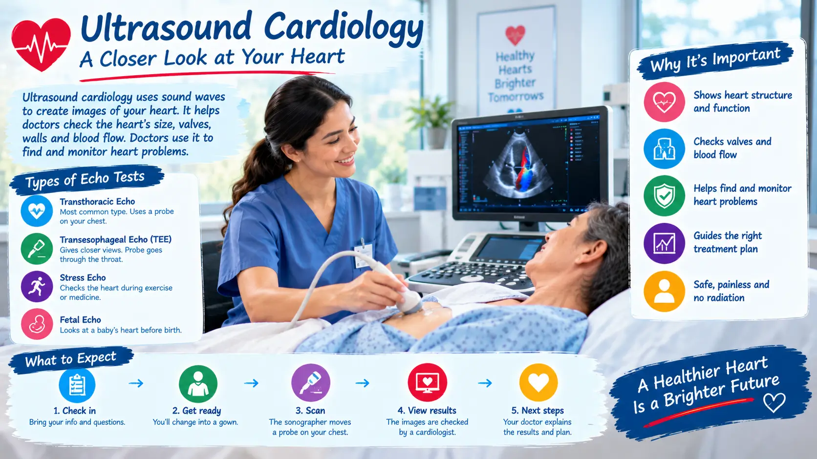

Ultrasound cardiology uses sound waves to create moving heart pictures. It helps doctors see heart size, valves, walls, and blood flow. Doctors use it to find and monitor heart problems.
A doctor may suggest a heart scan after a checkup or new symptoms. You may wonder what the test shows. You may also wonder if it hurts or needs special preparation. These are common questions.
Ultrasound cardiology usually means echocardiography, also called an echo. This test uses sound waves instead of X-rays. It can show the heart while it beats and pumps blood.
The test can check heart valves, chambers, muscle movement, and blood flow. Doctors may use different echo types for different needs. A standard echo looks through the chest from outside. Other types give closer views or show the heart during exercise.
This guide explains why doctors order an echo and what happens during it. You will also learn about cost, safety, results, and common echo types. Simple examples can help you know what to expect.
What Is Ultrasound Cardiology?
Ultrasound cardiology is heart imaging that uses sound waves. The medical name is echocardiography, or an echocardiogram. Many people simply call it an echo or heart ultrasound.
A small device sends sound waves toward your heart. The waves bounce back from heart structures. A computer turns those signals into moving heart pictures.
What Can a Heart Ultrasound Show?
An echo can show several parts of heart health.
- Heart size: It can show if chambers look larger than expected.
- Heart movement: It can show how the heart muscle moves.
- Pumping strength: It can estimate how well the heart pumps blood.
- Heart valves: It can show how valves open and close.
- Blood flow: Doppler imaging can show blood moving through the heart.
- Heart lining: It can show the sac around the heart.
- Some birth defects: It can show certain changes present from birth.
A standard transthoracic echo uses a probe on the chest. The word “transthoracic” means through the chest. It is the most common type of echo.
The American Heart Association lists echo as a common heart test. The American Society of Echocardiography also describes it as radiation-free imaging.
Why Would a Doctor Order an Ultrasound of the Heart?
Doctors may order an echo for many reasons. A heart murmur is one common reason. A murmur is an unusual sound heard through a stethoscope.
Doctors may also use an echo after chest symptoms or a heart event. The test can help assess valves and heart pumping. It can also help check some heart problems present at birth.
An echo does not give every answer. Your doctor may order other tests based on your symptoms and results.
Why Might You Need a Heart Ultrasound?
Doctors choose an echo based on your symptoms, history, and exam. The test can answer questions about heart structure and movement.
Some people need an echo because they have symptoms. Others need one because a doctor found a possible heart problem.
Common Reasons for an Echo
Your doctor may suggest an echo when you have:
- A heart murmur
- Shortness of breath
- Swelling in the legs
- Chest discomfort
- Fainting or near-fainting
- Certain abnormal heart rhythms
- Known valve problems
- A history of some heart conditions
- A suspected heart problem present from birth
Doctors may also repeat an echo to monitor a known condition. A repeat test can show whether heart function has changed.
Who May Need Different Types of Echo?
Most people have a standard chest echo. Some situations need a different approach.
A stress echo checks the heart during exercise or with medicine. It can show changes that may appear when the heart works harder.
A transesophageal echo, or TEE, gives pictures from inside the body. A thin probe passes into the esophagus, which sits near the heart. Doctors may use TEE when they need clearer views.
A fetal echo checks an unborn baby’s heart. Doctors may use it when a heart problem needs closer review before birth.
Three-dimensional echo can give views from more angles. Doppler imaging can show blood flow and its direction.
These tests serve different purposes. Your care team chooses the type that fits your medical question.
A Simple Patient Example
James has ankle swelling and gets short of breath at night. His doctor orders an echo to check heart function.
The result may guide the next step or lead to other tests.
What Symptoms Can Lead to an Echocardiogram?
An echo can be part of a heart check when symptoms raise concern. Symptoms alone do not tell doctors the exact cause.
Symptoms Doctors May Consider
Common symptoms can include:
- Shortness of breath
- Chest discomfort
- Unusual tiredness
- Swollen ankles or legs
- Fainting
- Fast or uneven heartbeats
- Reduced ability to exercise
- New or changing heart sounds
Some people have no symptoms. A doctor may still order an echo after an exam or other test.
An echo may also help monitor someone with a known heart condition. Repeat scans can show changes in heart function.
Not every symptom needs an echo. Doctors consider your history, exam, and other results.
Symptoms That Need Urgent Attention
Some heart symptoms need urgent medical help. Severe chest pressure can signal a heart attack. Trouble breathing can also be an emergency.
The American Heart Association advises calling 911 for heart attack warning signs. These can include chest discomfort, shortness of breath, sweating, or nausea. Pain may spread to the arm, back, neck, jaw, or stomach.
Do not wait for an echo appointment during a possible heart attack. Emergency teams can start urgent care and testing right away.
How Is an Echocardiogram Done?
A standard echo is usually simple and painless. A trained sonographer performs the scan in many care settings.
What Happens During a Standard Echo?
First, you usually change into a gown. You lie on a bed, often on your left side.
The sonographer places small sticky sensors on your chest when needed. These sensors can track your heartbeat during the test.
Next, the sonographer puts gel on a handheld probe. The probe moves over different areas of your chest.
The probe sends sound waves into your body. The returning waves create pictures on a screen.
You may hear machine sounds or feel gentle probe pressure.
Most standard echoes take less than an hour. Many people return to normal activities afterward.
Is an Echocardiogram an Ultrasound?
Yes. An echocardiogram is a type of ultrasound test for the heart.
Both use sound waves to create images. The main difference is the body area being examined.
An abdominal ultrasound examines belly organs. An echo focuses on the heart and nearby structures.
Echocardiogram vs EKG
An echo and EKG are different tests. They answer different questions.
| Test | Main purpose | What it shows |
| Echocardiogram | Checks heart structure | Chambers, valves, motion, and blood flow |
| EKG | Checks heart electricity | Rhythm, rate, and electrical patterns |
| Stress echo | Checks heart under stress | Heart movement during exercise or medicine |
Doctors may use both during one heart evaluation.
What About an Echo for Women?
Echo procedures are similar for both sexes. You may need to change into a gown for a chest echo.
The sonographer places gel on your chest and moves the probe over your skin. The scan does not use X-rays.
Ask about privacy, positioning, or a chaperone before the test.
What Do Echocardiogram Results Mean?
A heart ultrasound gives doctors information about structure and function. A doctor trained in echo reviews the images and prepares a report.
What Can the Results Show?
The report may describe:
- Heart chamber size
- Heart wall movement
- Pumping strength
- Valve opening and closing
- Blood flow
- Fluid around the heart
- Some structural changes
You may see a number called ejection fraction, or EF. EF estimates how much blood the left ventricle pumps with each beat.
A normal-looking result does not rule out every heart problem. An abnormal result does not always mean severe disease.
Your doctor combines the echo with your symptoms and other tests.
What If the Images Are Hard to See?
Sometimes body tissue, ribs, or lungs can make images less clear. The sonographer may try different positions or views.
In some cases, a small amount of contrast may improve the images. Contrast is a substance that helps certain areas stand out.
Tell the care team about allergies and medicines before contrast is used.
How Much Does an Echo Test Cost?
Echo test prices vary across the United States. Your final cost depends on the facility, insurance, test type, and billing rules.
A standard echo may cost less than a specialized echo. A TEE or stress echo can involve more staff and preparation.
Before the test, ask the clinic about the estimated price. You can also ask your insurer about your deductible and copay.
Ask whether the facility is in your insurance network. This can help you understand your likely out-of-pocket cost.
How Is a Heart Ultrasound Managed After the Test?
An echo itself usually does not need treatment afterward. The next step depends on what the images show and why the test was ordered.
What Happens After a Standard Echo?
After a standard echo, you can usually return to your normal routine. The report goes to the ordering clinician.
Your doctor may explain the results during a visit or by another approved method. Ask questions if any term feels unclear.
More testing may follow if the images raise a new question.
For example, Sara has a known valve problem. Her repeat echo shows stable valve movement. Her cardiologist may continue planned follow-up.
What About Stress Echo Results?
A stress echo compares heart images before and after stress. Stress can come from exercise or medicine.
Doctors look for changes in heart movement during stress. They also consider symptoms and other findings.
What About a TEE?
TEE can provide closer images of some heart structures and valves.
TEE needs more preparation because the probe passes through the throat. Medicine can help you relax and numb your throat.
You usually need someone to drive you home after sedation. Follow the exact food, drink, and medicine instructions from your care team.
How Can You Prepare for Better Heart Care?
Good preparation can make your visit easier. Most standard echoes need little special preparation.
Before a Standard Echo
Ask your clinic about your usual medicines. Most standard echoes allow normal food and drinks.
Wear comfortable clothing. Bring your medicine list and insurance information if requested.
Ye short versions try kar
Write down questions before the visit.
For Learners and Cardiology Students
Students may study echocardiography in ultrasound or cardiology programs. Training often includes anatomy, image views, patient safety, and scanning skills.
Heart ultrasound training can cover Doppler basics, heart measurements, lab practice, and clinical work.
Check school requirements before choosing a program.
The American Society of Echocardiography provides professional resources for cardiovascular ultrasound. Accredited labs may also follow quality standards.
A Caregiver Example
Lisa drives her father to an echo visit. She brings his medicine list and insurance card.
She also writes down his main symptoms. This helps the visit stay focused and organized.
When Should You See a Doctor About Heart Symptoms?
An echo is a useful test, but urgent symptoms need emergency care.
Call 911 for Emergency Symptoms
Call 911 for possible heart attack signs. Do not drive yourself to the hospital.
Emergency warning signs can include:
- Chest pressure, squeezing, or fullness
- Trouble breathing
- Cold sweat
- Nausea or vomiting
- Fainting or severe weakness
- Pain in the arm, back, neck, jaw, or stomach
Women and older adults may have less typical symptoms. Severe or sudden symptoms still need urgent attention.
The American Heart Association recommends emergency care for suspected heart attack symptoms.
When to Arrange a Regular Visit
Make a medical appointment for new or ongoing symptoms. These can include shortness of breath, swelling, fainting, or unusual tiredness.
A doctor can decide whether an echo may help. Other checks may also be needed.
Do not order an echo on your own based on symptoms alone. The right test depends on your health needs.
Questions to Ask Your Doctor
Consider asking:
- Why do I need this heart ultrasound?
- Which type of echo will I have?
- Do I need to change my medicines?
- Can I eat and drink before the test?
- When will I receive my results?
- What happens if the images are unclear?
- What will the next step be?
Clear questions can help you understand your care plan.
Frequently Asked Questions
Q: Is an echocardiogram an ultrasound?
A: Yes. An echocardiogram is an ultrasound of the heart. It uses sound waves to create moving heart images. It does not use X-rays or radiation.
Q: Why would a doctor order an ultrasound of the heart?
A: A doctor may order one to check heart structure and pumping. It can also examine valves, heart movement, and blood flow. The reason depends on your symptoms and medical history.
Q: How much does an echo test cost?
A: The price varies by location, insurance, and test type. Your clinic and insurer can give a better estimate. Ask about deductibles, copays, and network status.
Q: Is an echocardiogram painful for women?
A: Usually, no. A sonographer moves a gel-covered probe across the chest. You may feel mild pressure during some views.
Q: What is the difference between an echocardiogram and an EKG?
A: An echo shows heart structure, movement, valves, and blood flow. An EKG records the heart’s electrical activity. Doctors may use both tests for a fuller heart check.
Q: What does ultrasound cardiology mean for students?
A: It usually refers to learning heart ultrasound and echocardiography. Students may study anatomy, image views, Doppler, and patient care. Training rules depend on the school and program.
Q: Is an echo safe during pregnancy?
A: Echocardiography uses sound waves rather than ionizing radiation. Your doctor can decide which heart test fits your situation. Ask your care team about any test during pregnancy.
Conclusion
Ultrasound cardiology helps doctors see the heart while it beats. An echocardiogram can show heart size, movement, valves, and blood flow.
Most standard echoes are painless and need little preparation. Different echo types can answer different medical questions.
The test can monitor known heart problems or support a new heart evaluation.
If you have new or ongoing heart symptoms, ask your doctor about your options. Seek emergency help for possible heart attack signs.
Talk to a cardiologist if you have heart concerns.
Medically reviewed for accuracy. Always consult a licensed cardiologist for personal advice.